


 Shopping Cart
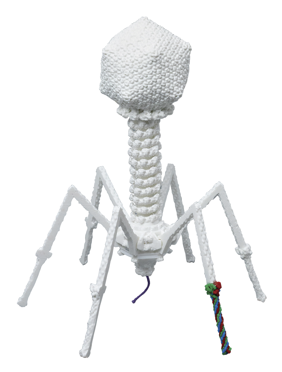
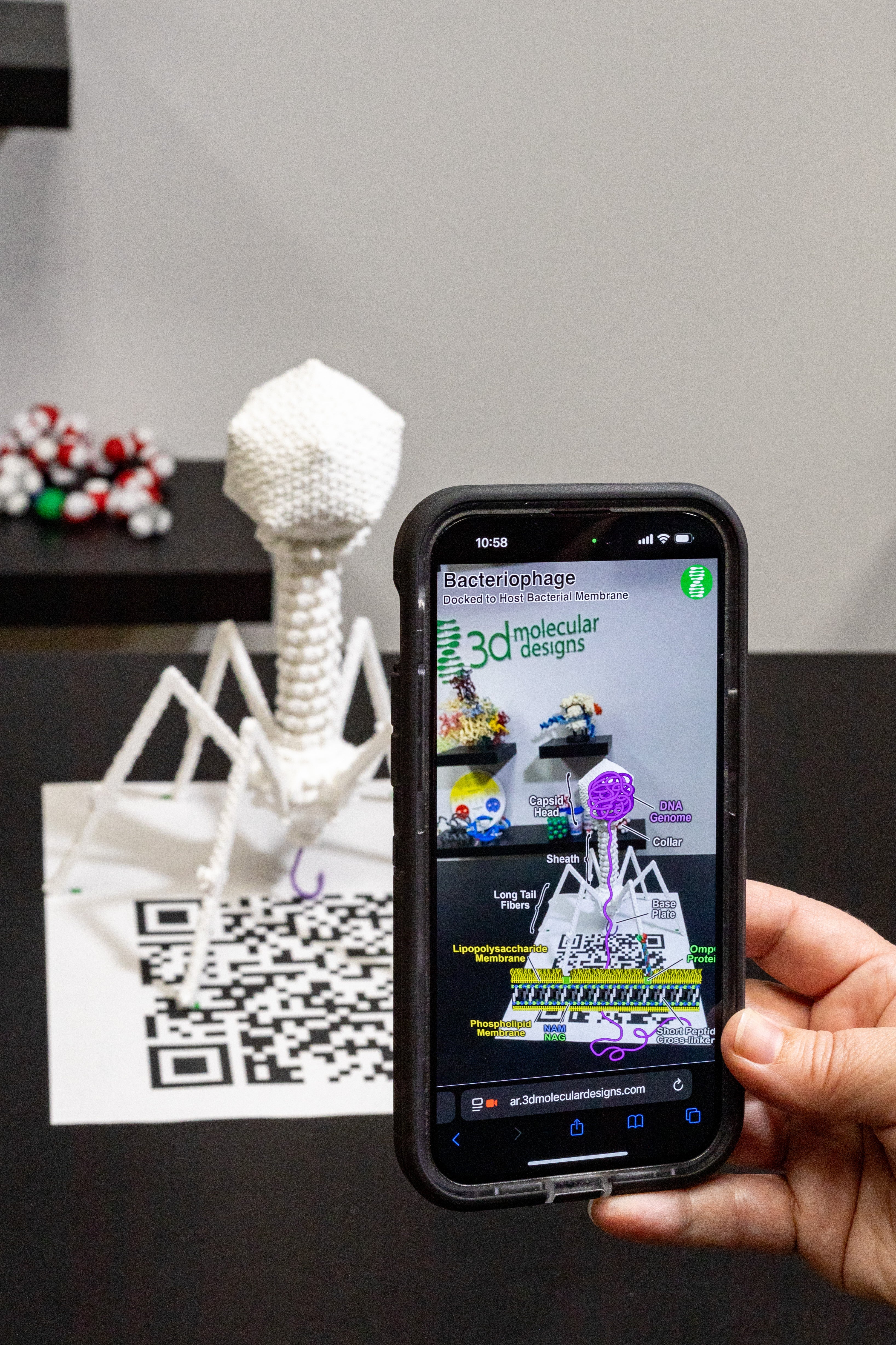


T4 Bacteriophage Mighty Model©
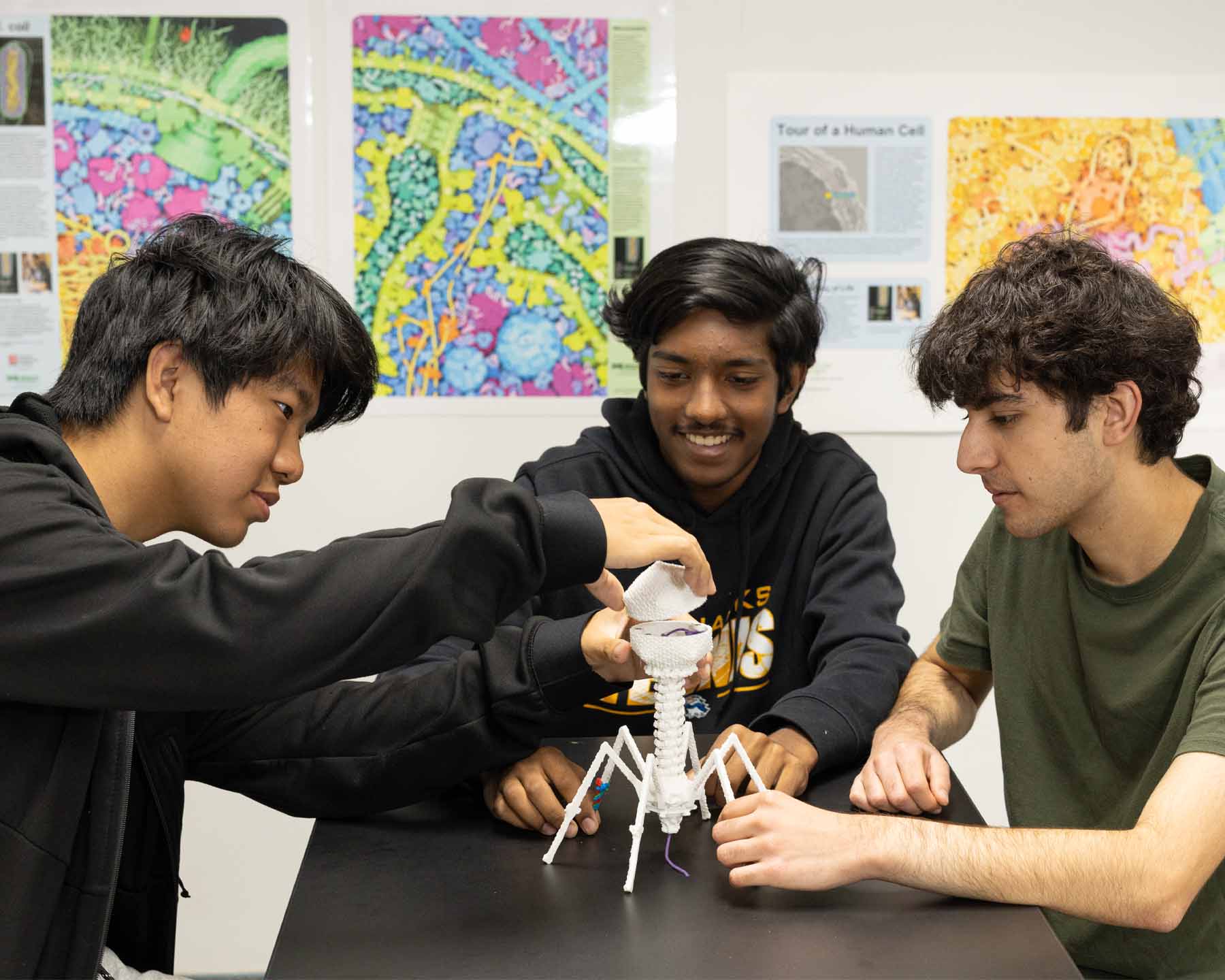
As your students launch into modeling the ongoing battle of survival between T4 bacteriophage and E. coli, they‘ll first encounter the amazing structure of T4 bacteriophage before exploring new membranes, and an assembly form of reproduction. Armed with two slide decks, you’ll guide your students, as they:
-
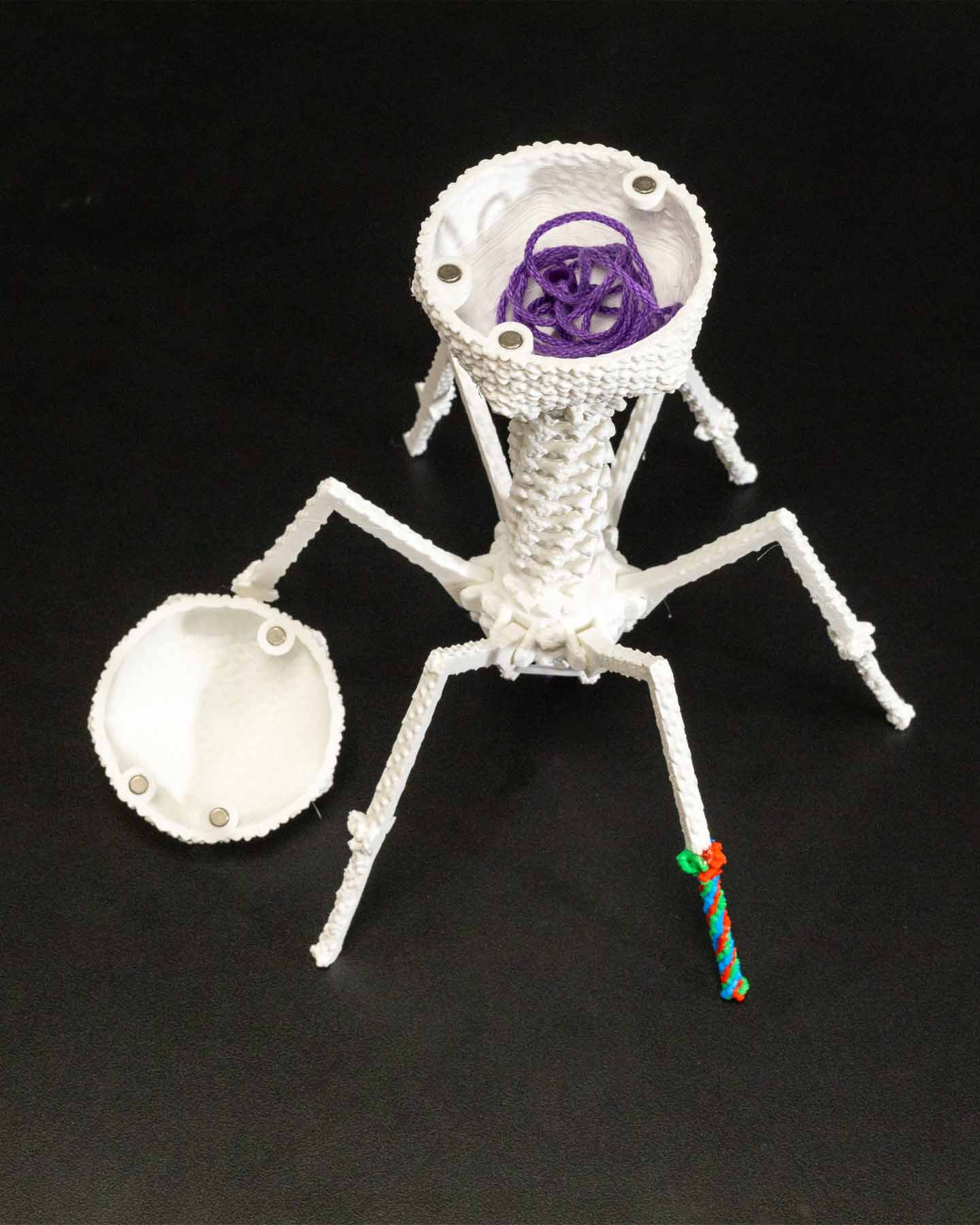
Identify the parts of T4 bacteriophage, which is comprised of an astounding 2,000 proteins
-
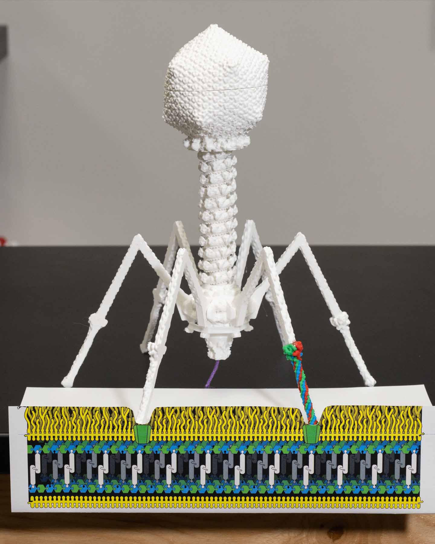
Predict how the T4 phage infects E. coli, using the T4 model and a printable E. coli membrane graphic
-
Explore the Gram-negative E. coli double membrane and contrast with a Gram-positive Staphyloccoccal single membrane.
-
Model the four phases of a T4 phage infection
-
Discover how T4 destroys E. coli’s DNA before replicating, transcribing and translating its own DNA by co-opting E. coli’s mechanisms for these processes.
-
Compare and contrast lytic and lysogenic viral life cycles
-
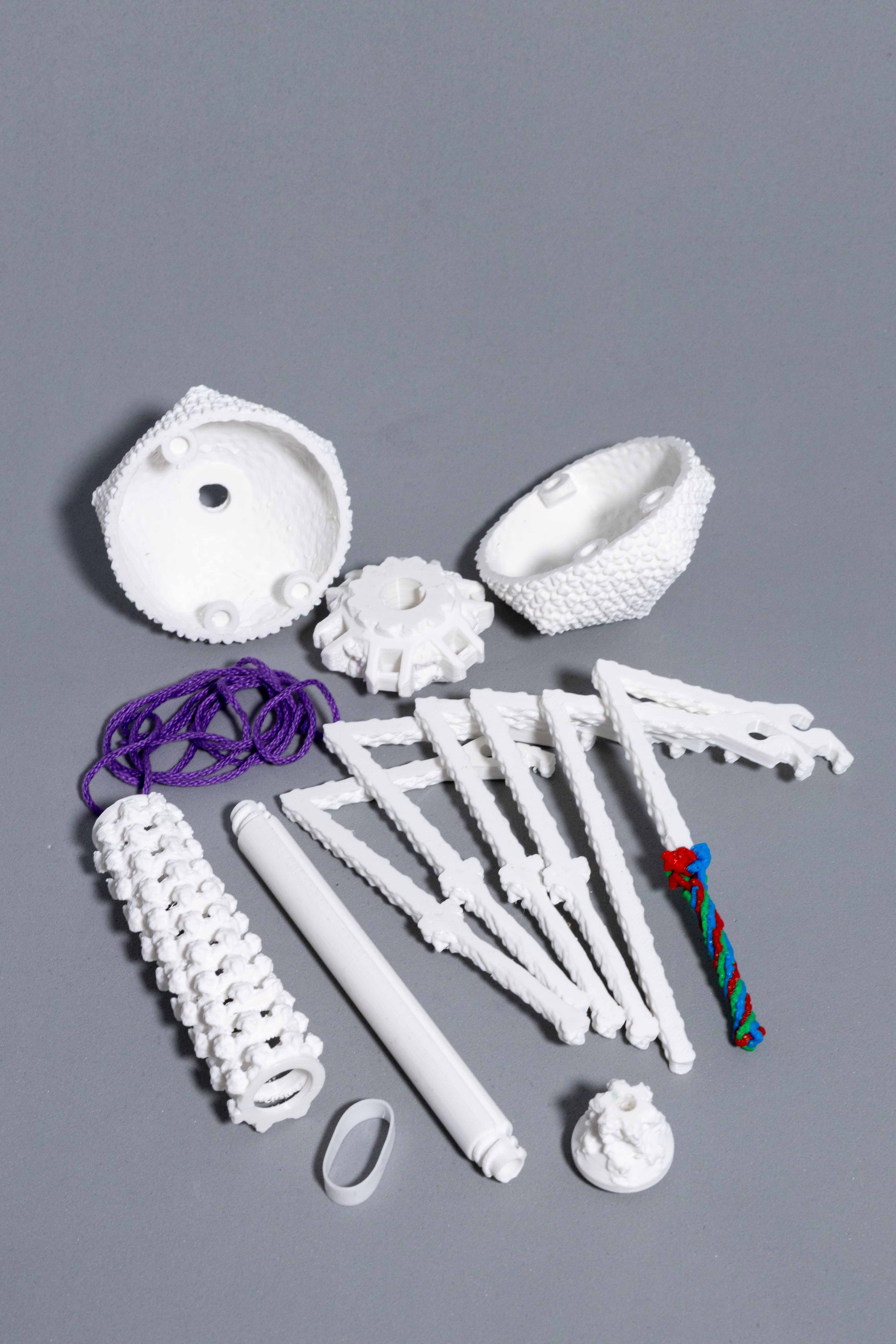
Assemble their T4 bacteriophage model, as assembly would occur in E. coli during virus replication
-
Discover how all the newly produced T4 bacteriophages break out of their E. coli host
While every part of T4 bacteriophage provides a structure-function story, you’ll be able to differentiate use of the model by challenging your advanced students to discover how T4’s spindly-looking legs (long tail fibers) can be strong enough to support its oversized head and complex center tail mechanism. By examining its gp37 amino acid sequence – at the tip of the long tail fiber – they may discover the answer in its unique pattern. With your subscription to our Digital Modeling Hub, they can further explore the structure.
